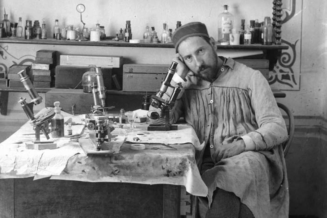
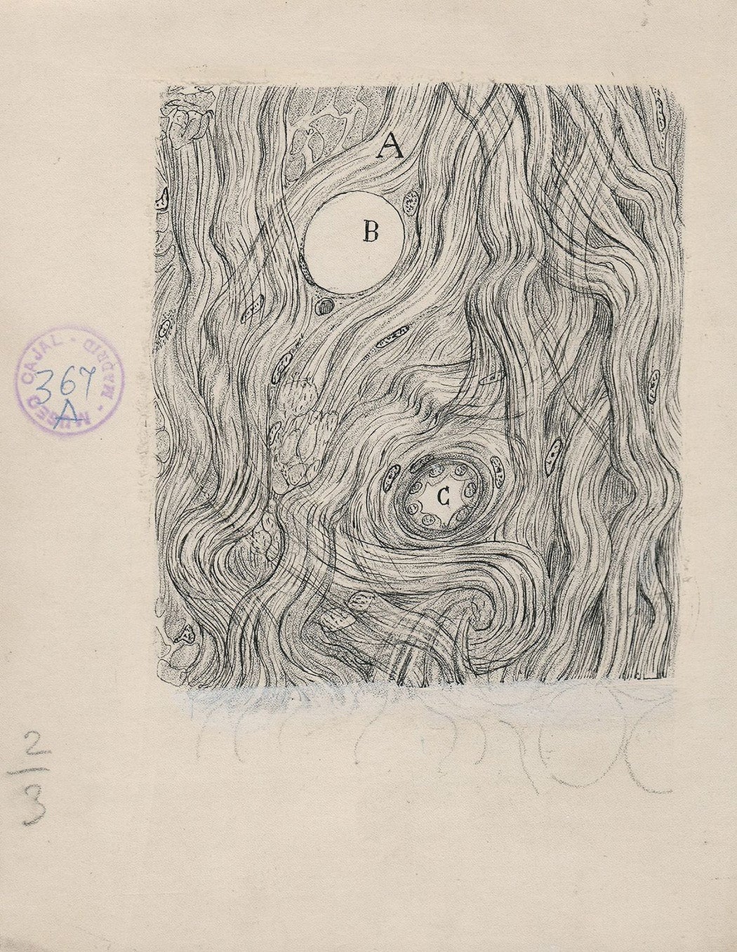
In 1887, neuroscientist Santiago Ramón y Cajal visited Madrid, where he viewed microscope slides of nerve cells prepared with a relatively new technique, the “black reaction,” for the first time.


“Against a clear background stood black threadlets, some slender and smooth, some thick and thorny, in a pattern punctuated by small dense spots, stellate or fusiform. All was sharp as a sketch with Chinese ink on transparent Japan-paper,” he recounted, “Dumbfounded, I could not take my eye from the microscope.”
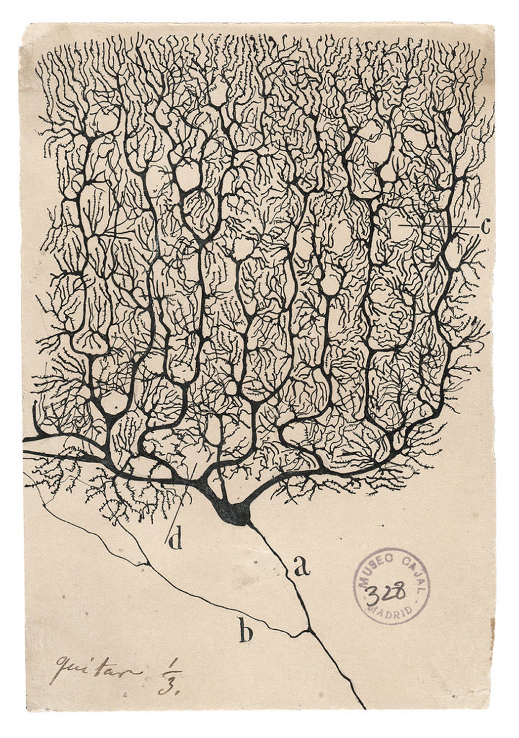
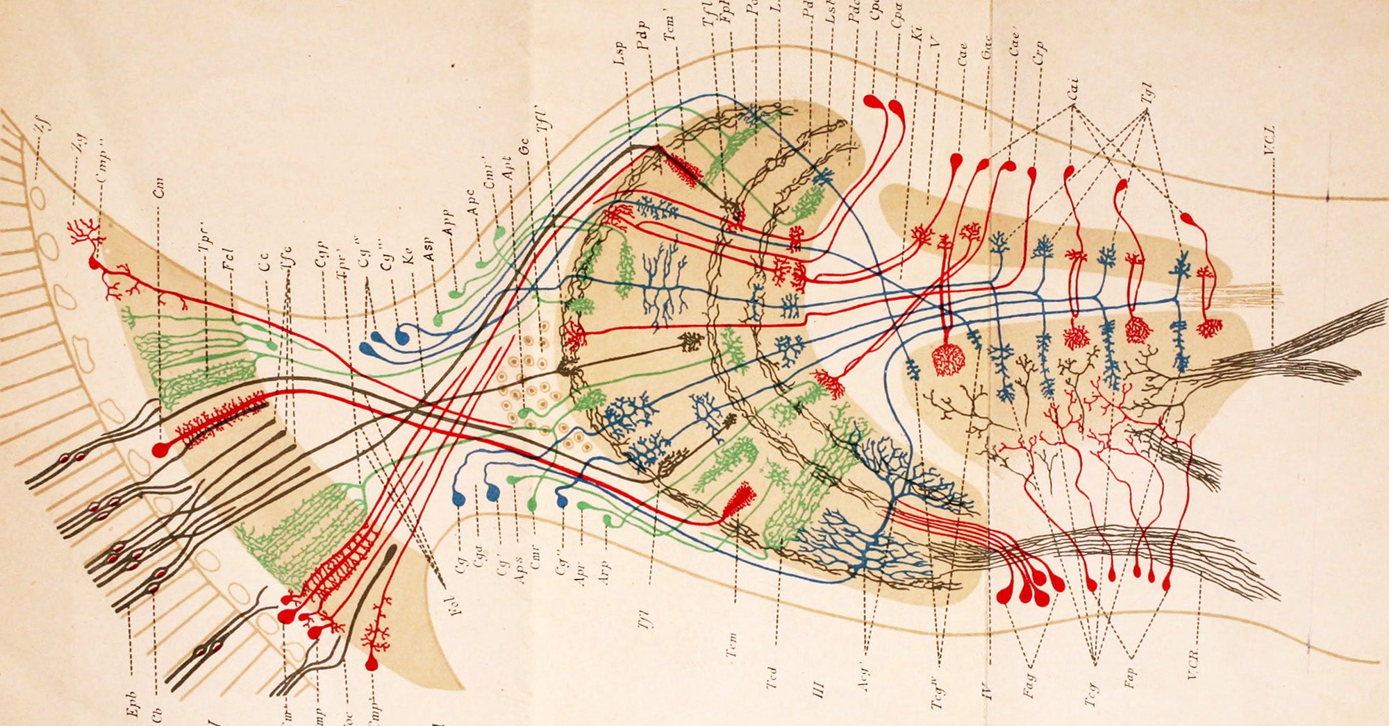
Although Cajal would go on to use other staining methods in his scientific investigations, he is inextricably tied to the black reaction and its originator, Camillo Golgi. The two shared a 1906 Nobel Prize for understanding the structure of the nervous system. Yet they were on opposing sides of a scientific dispute. Golgi and conventional thought held that the brain was a vast, connected net of nerves: the reticular theory. Cajal found that neurons were individual cells, communicating between branching tips at small gaps, or synapses.
This neuron doctrine, as it came to be called, is one part of Cajal’s lasting influence, which broadly speaks to the anatomy, structure, and function of the brain and nervous system. All of it is at least in part a product of, and arguably not possible without the remarkable, delicate drawings Cajal made to illustrate his observations of the brain and extended nervous system.
“I think cellular neuroscience today is still standing firmly on the images that Cajal generated at the turn of the twentieth century,” said Marina Picciotto, a neuroscientist at Yale, “It’s amazing how many neuroscience talks I go to where one of the first slides is an image from Cajal.” Picciotto, the former editor in chief of the Journal of Neuroscience, is president-elect of the Society for Neuroscience.
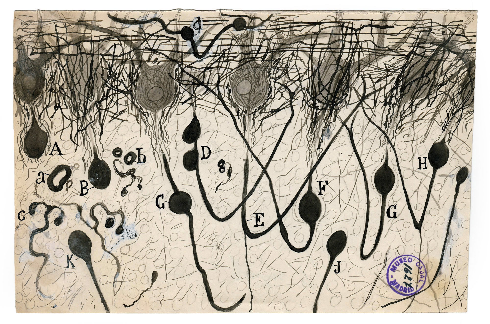
Cajal’s drawings of neurons are among the most important images in the field. He’s remembered as a masterful artist, but abbreviated biographies often omit the fact that he was also an avid photographer. For Cajal, drawing was a choice, motivated not only by deep knowledge of both the potential and constraints of contemporary photographic techniques, but a belief in the importance of the trained artistic hand and scientific eye.
Photographers had experimented with microscopes since the early days of the medium. So photomicrography, albeit with technical limitations, was available to Cajal at the time of his first historic observations in the late 1880s.
For example, in 1839, the pioneering photographer William Henry Fox Talbot presented “Some account of the Art of Photogenic Drawing, or the Process by which Natural Objects may be made to delineate themselves without the aid of the Artist’s Pencil.” In his remarks, Talbot mentioned the use of a solar microscope, a device which could project an enlarged image of an object onto a viewing surface or photosensitive paper. As early (or late) as 1867, a doctor named Richard Maddox was arguing for the practical case for photomicrography in medicine and other areas.
For Cajal’s purposes, photography’s limitation with respect to understanding neurons boiled down to a flattening effect, said Susan Goetz Zwirn, a professor of Art Education at Hofstra University.
“He wanted to explore multiple focal points in his depiction of the neuron,” both over the surface of the slide and through the depth of the tissue, said Zwirn. “He felt this could only be accomplished through drawing with a variety of artistic media.” Microscopes could only bring a relatively narrow, shallow section of a slide into focus at a given moment, which was all a camera was capable of capturing.
Cajal could survey a section of tissue and then, with pencil and ink, depict fine detail across the entirety of a cell, and convey depth through variation of line thickness and materials. Doing so required not just microscopy and artistic skill, but knowledge and insight about what to look for. “At that time there was no other way to explain his results,” said Virginia García-Marín, a neuroscientist at York College, who has worked with Cajal’s archive.
Furthermore, drawing was fundamental to Cajal’s science, according to Zwirn. If drawing is a way of looking at things, without the artistic techniques, Cajal’s observations could have been very different and limited. “He said that all neuroanatomists must be artists,” said Zwirn. “He doesn’t see how anyone could be one without being an artist.”
Drawing can still be an important laboratory skill, for example, for science students trying to make sense of what they see under the microscope, said García-Marín. “You have to draw, or you aren’t going to be able to process all this information.”
Cajal’s views on the potential advantages of photography, particularly the perceived objectivity, are difficult to pin down—perhaps as difficult as defining scientific objectivity itself. “It is well-known that man projects his personality onto everything, and that when he believes he is photographing the outside world he is often observing and depicting himself,” Cajal once wrote. He also recommended including photomicrographs with words and illustrations, which “guarantees the objectivity of our description.”
One benefit of scientific photography may be that many steps are mechanically repeatable in a way that drawing is not, using similar equipment, settings and processes at a different time or by a different person. But if one of the main differences between a camera lens and human vision is objectivity, it may come with drawbacks. “[Photography is] very limiting,” said Zwirn, “It’s not capturing the full potential and scope of the human eye.”
The Golgi method presented a clear picture of the gross anatomy of the neuron but didn’t allow for investigation of the insides of the cell. As his research questions evolved, Cajal needed new techniques to prepare his slides. Throughout his career he experimented with ways of staining brain tissue, many of which involved chemicals also used in photographic processes, such as potassium dichromate, silver nitrate, and gold chloride. His photographic background benefited his scientific endeavors even when he wasn’t taking pictures. These new histological techniques were accompanied by new artistic techniques, whether he used pencil and ink, or added watercolors.
Cajal’s Royal Society obituary, written by the Nobel prize-winning physiologist Charles Sherrington, who coined the term “synapse,” mentions photography only as a “pastime,” although it notes that label is questionable. Since childhood, photography had been among Cajal’s many interests or sidelines, which reportedly also included bird-watching, chess, literature, writing science fiction, and, of course, sketching. He wrote a book on color photography, and when Spain’s Royal Photographic Society was founded in 1899, was named honorary president.
Later in life, Cajal investigated various photographic methods, such as stacked exposures, stereographs, and moving pictures for capturing the same information he wanted his illustrations to communicate. New technologies have made some of these processes possible or much easier than they would have been a century ago. For example, computers can automatically stitch together many microscopic images into a single mosaic with many focal points.
And modern neuroscientists have many options besides microscopic photography for producing images of the nervous system or monitoring its activity. Two important methods, computed tomography (CT) and magnetic resonance imaging (MRI), are around fifty years old today, the same age as the daguerreotype and early photomicrographs when Cajal first viewed the black reaction. García-Marín has revisited Cajal’s slides, photographing them and even producing three-dimensional computer reconstructions.
Despite these advances, experts said that Cajal’s general approach to images is still a great example to scientists. “He was not only looking at these images as something static,” said García-Marín, “He was able to see physiology, the function—what does it mean for the neuron?”
“I think one of the primary functions of our brains is to make coherent stories from disparate pieces of information,” said Picciotto, “Interpreting disjointed images into a functional whole, as Cajal did in so many of his images of neurons or brain areas, is like telling a story after hearing many different jumbled viewpoints.”
Cajal’s heralded artistic abilities were important to his science; his photography perhaps less obviously so, in ways which underscore the value he invested in the drawing process. Taken together, there’s much to consider about how to look at images, how to produce them, and how to communicate about them.
Weekly Newsletter
"*" indicates required fields
While many pieces of Cajal’s writings still seem valuable or forward-looking, others might feel outdated, or still unfortunately relevant to modern times. Cajal called a few women associates, but his close scientific circle was mostly male. “For the man of science, the aid of a wife is just as necessary in youth as in old age. A woman at one’s side may be likened to a knapsack in battle: without the accessory one fights unencumbered, but after the battle, then what?” he wrote in his Advice for a Young Investigator.
García-Marín quotes the same book at the bottom of her emails, replacing the original “hombre” or “man” with “ser humano” or “human being.”
“Todo ser humano, si se lo propone, puede ser escultor de su propio cerebro.”
“Any human being could, if so inclined, be the sculptor of their own brain.”
.jpg)
_(20675334682).jpg)