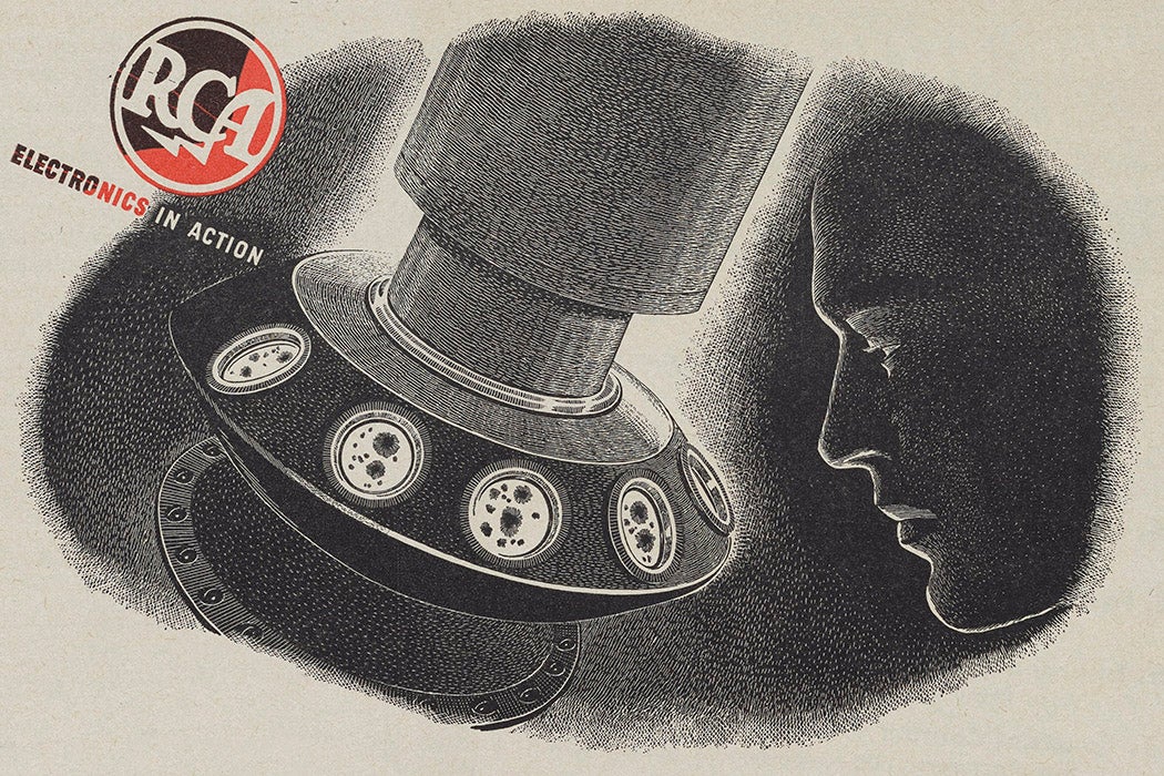
As flu season approaches, it’s a good time to look back at the invention that made virology possible. Viruses weren’t well understood until somewhat recently, in part because they couldn’t be seen even under a microscope. Historians Ton Van Helvoort and Neeraja Sankaran argue that the invention of the electron microscope initiated a paradigm shift by making viruses visible. They begin their story with a very curious scientific presentation.


In 1942, Ladislaus Marton gave a lecture at Stanford titled “Alice in Electronland.” The physicist presented it as “an original manuscript of Lewis Carrol” that he found hidden in a bookstore. In this unconventional lecture, Alice looks at some strange photographs, then shrinks so small that she can’t even see her own fingers.
Marton uses the story to explain how the newly perfected electron microscope worked. Alice can’t see her own fingers because they’re smaller than the wavelength of visible light. This makes them “submicroscopic,” meaning that a conventional microscope couldn’t reveal them. An electron beam with a much smaller wavelength, however, could be used to produce an image of them.
To see things this small, scientists couldn’t put samples on glass plates like they did for normal microscopes. Glass was too thick and would have deflected and absorbed too many electrons. Even air would have interfered with the electron beams, so the imaging had to be done in vacuum.
Researchers put virus samples into a collodion that was spread thinly on a fine mesh. Electrons shot through the gaps in the mesh toward a photographic plate, forming images of the viruses suspended within the gaps. With these techniques, write Helvoort and Sankaran, scientists “discovered facts that altered the very definition of the viruses.”
The limitations of conventional microscopes meant that before the electron microscope, “virus” was a generalized term—researchers knew that submicroscopic organisms existed, mainly because of the diseases they caused. Facing a lack of direct knowledge, biologists originally classified viruses by the type of disease they caused or the type of host they infected. But micrographs revealed their size, shape, and structure, and scientists began to understand a little more about how they worked and how they might be related to one another. It became clear that viruses were distinct from bacteria and were in a category by themselves.
Weekly Newsletter
The tobacco mosaic virus was one of the first that was studied intensively. Soon scientists imaged many others, including influenza. Earlier studies of influenza were limited, write van Helvoort and Sankaran—it was the electron microscope that allowed researchers to make “definitive claims about various peculiarities of structure and the eclipse period of the influenza virus.”
Scientists had also used X-ray crystallography to image viruses early on. But van Helvoort and Sankaran believe that the detail contained in micrographs gave a more useful view of “Electronland.” These images became both “evidence and arguments” that redefined the nature of viruses and began a conversation about how to classify and study them.
Teaching Tips
- J. M. C., “Mosaic Disease of Tobacco” (1922)
- Edwin E. Slosson, “Starting a New Disease” (1925)
- B. T. Dickson, “Tobacco and Tomato Mosaic” (1925)
- W. C. Price, “Local Lesions on Bean Leaves Inoculated with Tobacco Mosaic Virus” (1930)
- W. M. Stanley, “Isolation of a Crystalline Protein Possessing the Properties of Tobacco-Mosaic Virus” (1935)

